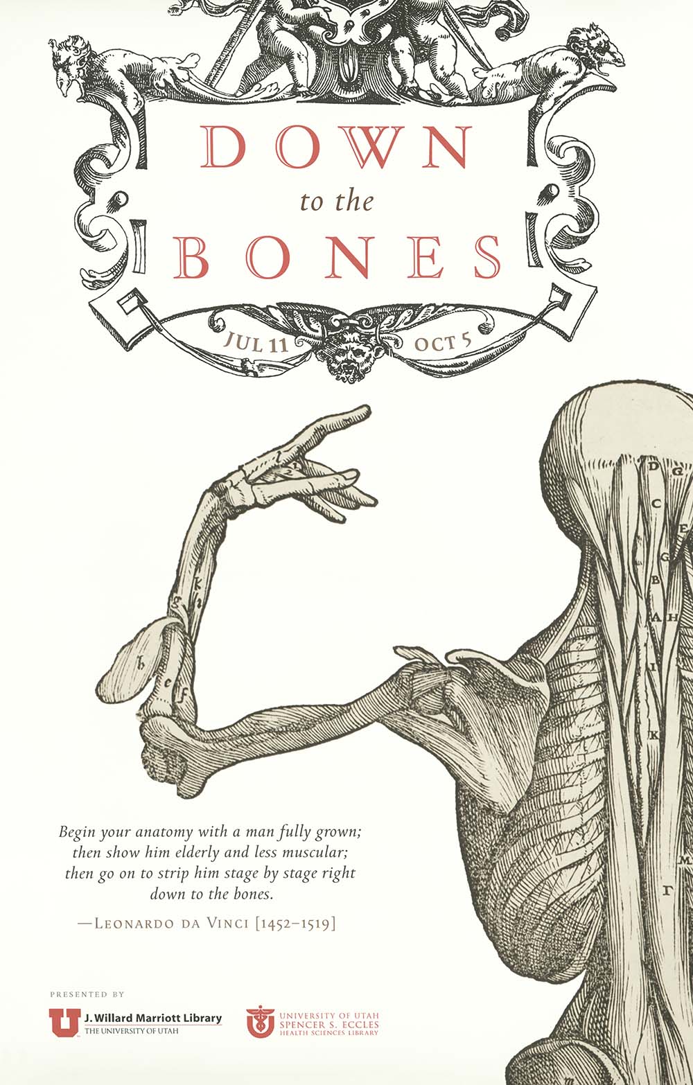
Down to the Bones
"Begin your anatomy with a man fully grown; then show him elderly and less muscular; then go on to strip him stage by stage right down to the bones." -- Leonardo da Vinci (1452-1519)
Checklist for "Down to the Bones"
Curated by Luise Poulton, 2014
Digital exhibition produced by Alison Elbrader, 2016
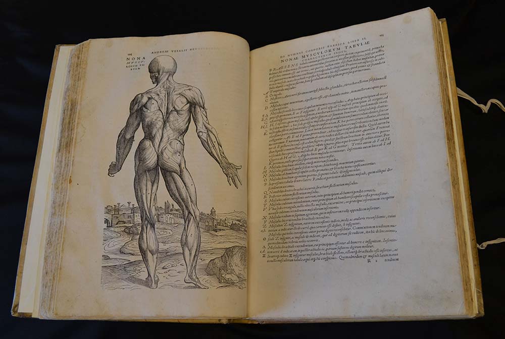
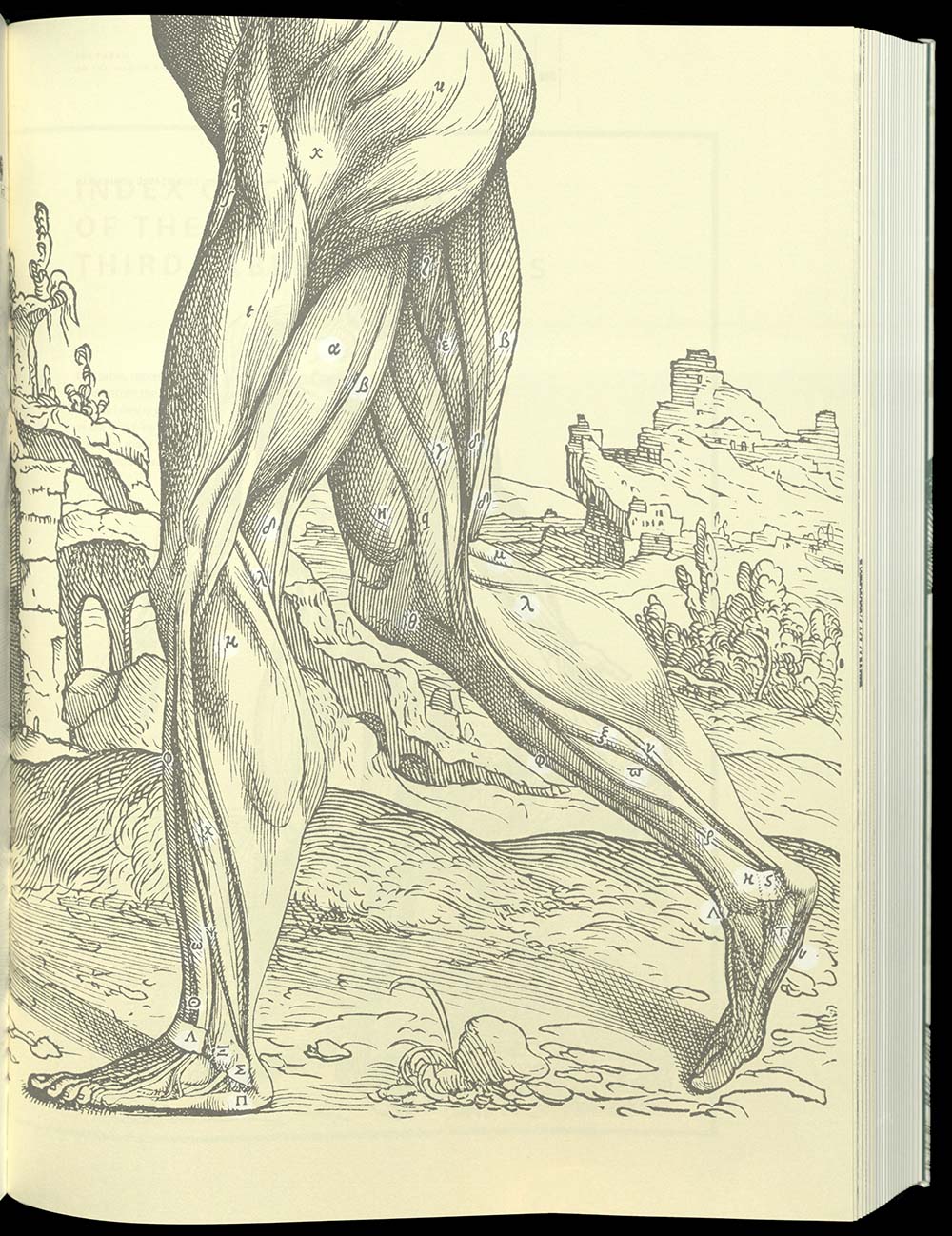
De Humani Corporis Fabrica of Andrea Vesalius (1514-1564) was an exquisite piece of creativity that blended observation; organization of information, format, typography; and illustration into an integrated whole to accurately describe the human body. The intense collaboration between scientist, artist, and printer was unprecedented. Prior to the publication of this book, medical texts were mostly derived from the medieval Arabic medical tradition or from translations of the works of Classical authors, whose texts had been corrupted by translation and re-translation: from Greek into Syriac, Syriac into Arabic, Arabic into Latin. Renaissance Europe embraced the classical works of Hippocrates and the Greco-Roman Galen. Vesalius, however, chose to further his knowledge of human anatomy by studying human cadavers. From these studies, Vesalius formed his position that the validity of any hypothesis rested solely upon facts established by observational methodology. His work marked the beginnings of modern science.
First edition
This book was published in the same year that Copernicus published his heliocentric theory. Andreas Vesalius’ illustrated book on anatomy marked the beginnings of modern observational medicine. Detailed medical illustrations depict precise anatomical detail, and are fine examples of the naturalism of Italian Renaissance art, which was based on the newly discovered principle of linear perspective. A number of artists, including possibly the Venetian master Titian, created the drawings for the woodcuts which illustrate and relate precisely to the text. The sequence of illustration from skeleton to various muscle layers and nerves succeeded in clarifying the study of the human body to a degree never before achieved. The book was also a feat of typographical genius. Typefaces were introduced in the heading of Book I, including a cursive type – used for the legends of the illustrations and to a lesser extent for the marginal notes. The marginal notes are placed in the outside margins and the inside margins; the notes in the outer margins refer to the contents of the text, and those in the inner margins are cross-references to the illustrations. Ornamented capital letters introducing two hundred and eight chapters depict the art of dissection, surgery, and obstetrics. Fourteen muscle figures form a series of illustrations linked by a continuous background, an accurate panorama of an area west of Padua. The meticulous printer, Johannes Oporinus, had studied medicine, Latin, Greek and Hebrew. All three of these languages are used in Fabrica. On loan from the Spencer S. Eccles Health Sciences Library.

QM25 V4 1555
The second edition of Vesalius’ “Anatomy” is considered better than the first for its superior production. The paper was heavier, enabling clearer reproduction of the illustrations. A new typeface, designed by Parisian Claude Garamond after a classical Roman type from the Renaissance, was used for the text. For the 1555 edition, new and slightly larger initials were cut, a consequence of the adoption of a larger typeface. The original plates were slightly modified by cutting away the wood around some of the guide letters where they tended to be obscured by shadow. Vesalius added to the second edition corrections, additions and new information enabling the discovery of the circulating nature of blood. Vesalius extended his report on the effects of the nerve section, on laryngeal paralysis, on artificial respiration by intratracheal intubation, and the continuation of life after the removal of the spleen.

Facsimile
This text is one of the oldest extant and most important links between Greek classical and European medicine of the High Middle Ages. Abu al-Qasim was born in 936 AD at al-Zahra near Cordoba in Spain, during the flourishing of the Ummayad caliphate. He was the personal physician for the caliph al-Hakim II (961-976). His writings were widely known in his own time and preferred to those of Galen. Much of the medical knowledge from the classical world was lost to Western Europe by the seventh century. The Arabs reintroduced it to the West, but also improved on it. Chirurgia is the last chapter of a thirty volume work, Al-Tasrif, combining all of the then known Greek medical knowledge and the experiences of the physician himself. Only three sections of the entire work were translated into Latin, by Gherard of Cremona (1114-1187). Chirurgia includes information on cauterisation, midwifery, bloodletting, and bone setting. This fourteenth century copy is richly illustrated, underlining the high esteem in which it was held in the western world. Sixty-eight miniatures illustrating individual methods of medical treatment and instruments decorate the text. At the center of most of the miniatures is the dignified and noble physician, dressed in long, flowing robes. For the patients’ part, pain and fear can be read in their faces.
Facsimile
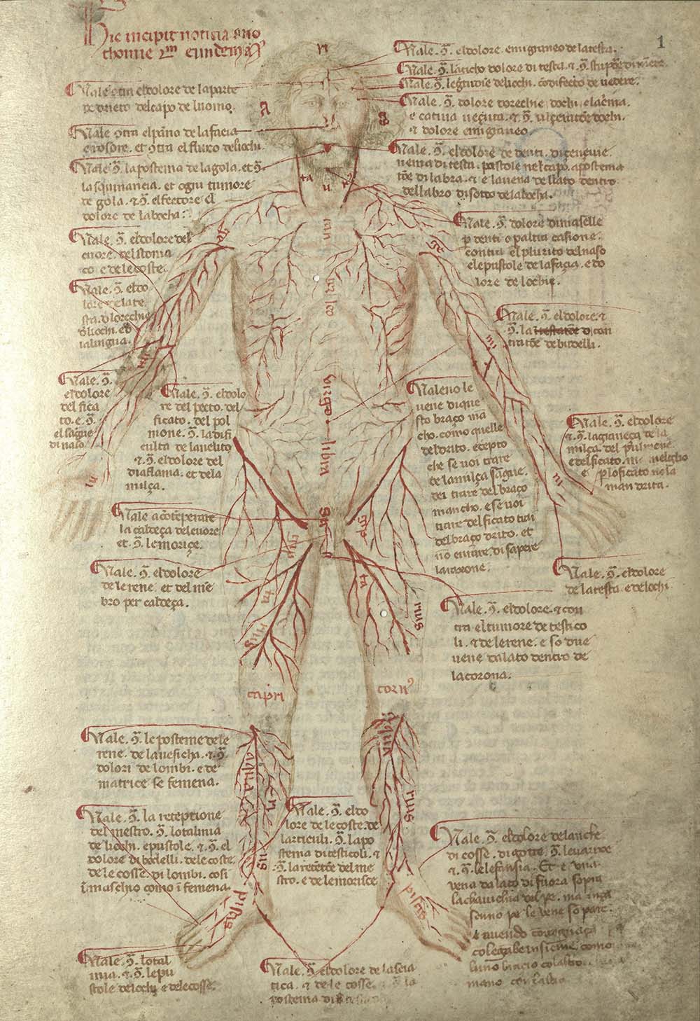
Bartolomeo Squarcialupi was a Paduan physician. His treatise describes the practice of cauterization, that is, the application of hot irons (cauteries) on body parts presenting a wide range of diseases. The drawings in this manuscript, by an unknown artist, reflect a Paduan pictorial tradition deeply influenced by Giotto. This volume is an assemblage of the remains of a larger manuscript. The assemblage likely took place in the 1800s and reflects a later organizational style, placing loose leaves near text and illustrations presenting similar content. Three separate medical texts are represented. Facsimile edition of one hundred and ninety-nine copies.
Facsimile
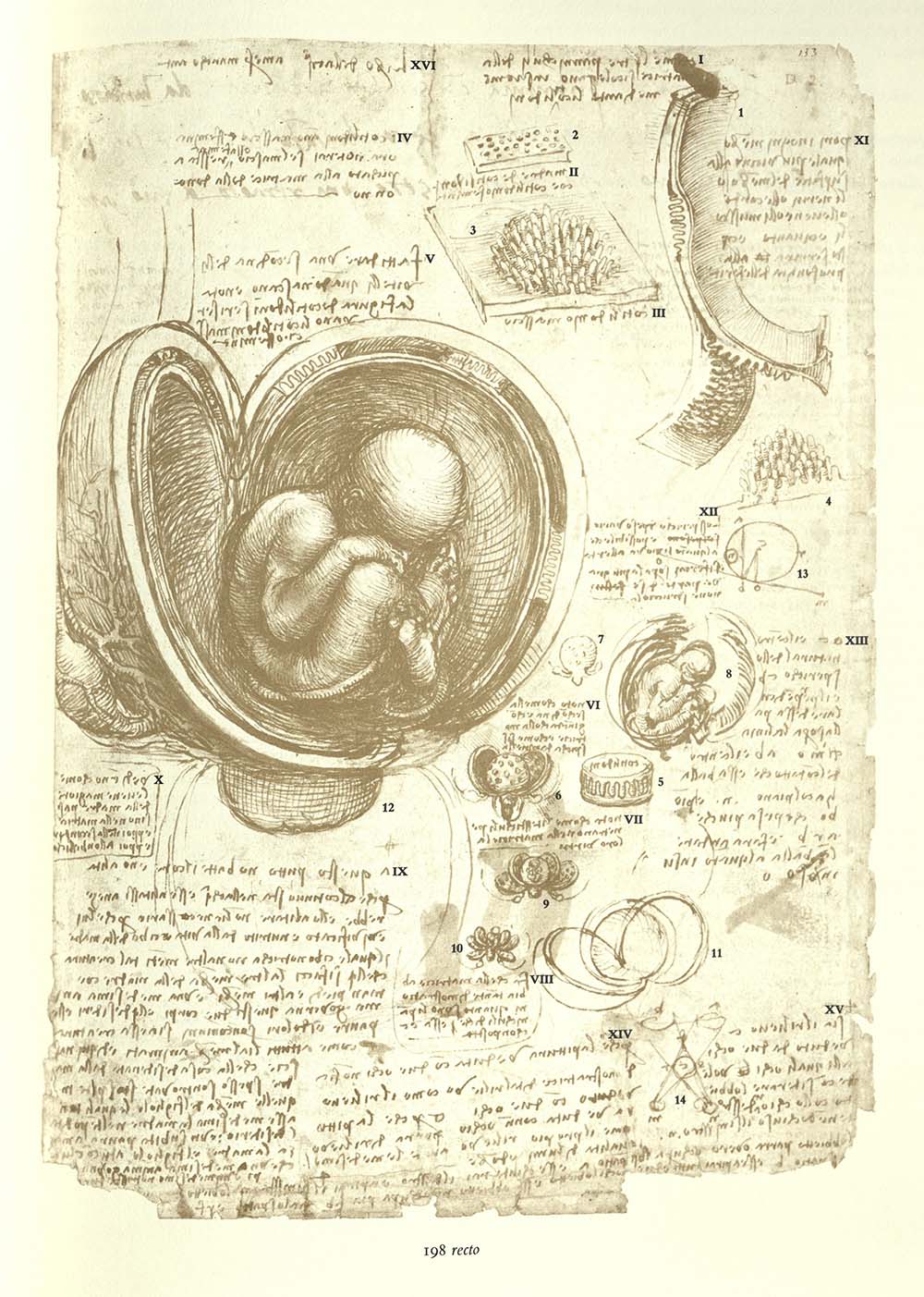
Over a period of twenty years Leonard performed more than thirty dissections of human cadavers. He made thousands of drawings of the human body, recording them on the pages of many notebooks. He intended to publish his treatise on anatomy but died before he was able to do so. Several artists made drawings from Leonardo’s studies, but, for the most part, his notes and drawings languished unknown within his private papers and went undiscovered for the next four hundred years. The contents of Leonardo’s studio were passed on to a pupil, and then to unknown hands. Many of the drawings were later bound into volumes and collected by the royal courts throughout Europe. It is unknown when this collection of anatomical drawings arrived at Windsor Castle. The earliest record of its existence notes that it was in the possession of Queen Mary II in 1690. The collection was re-discovered by physician William Hunter (1718-1783). This facsimile edition includes transcriptions, translations and anatomical notes by K. D. Keele and commentary on the reassembled sheets and their chronology by Carlo Pedretti. The facsimile consists of two text volumes and one portfolio box bound in Nigerian goatskin dyed royal blue. The spines are embossed in gold. Paper is specially-designed 100% rag mold-made paper, with gilded edges. Facsimile edition of nine hundred and ninety-eight copies. University of Utah copy is no. 48.
First German edition
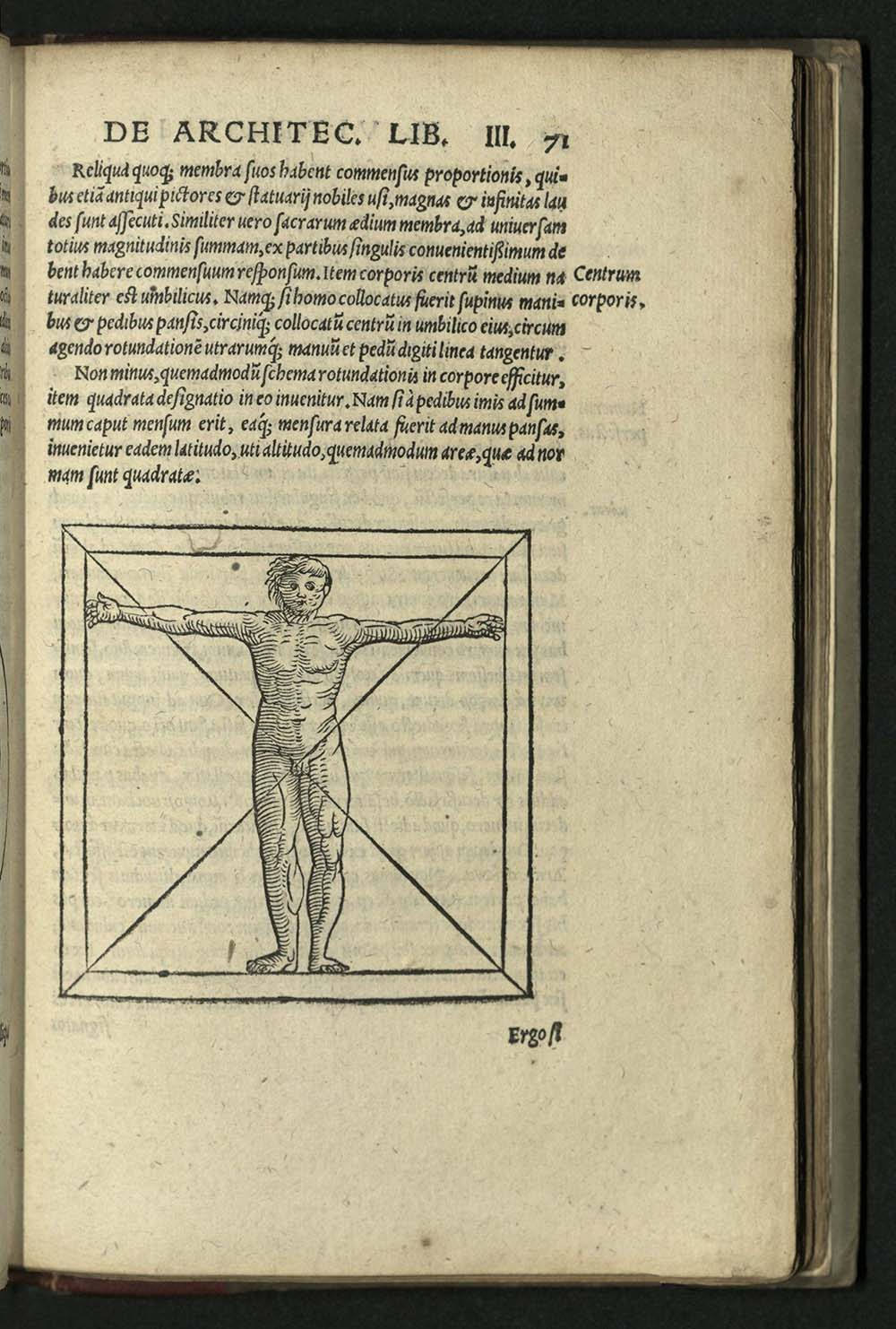
Pollio Vitruvius was a Roman architect. It is possible that he served as a soldier under Julius Caesar, most probably as an army engineer. His “Ten Books of Architecture” is the only surviving work on architecture from classical antiquity. The original illustrations had long been lost by the time the first printed edition was produced. New illustrations were added to the sixteenth century editions, beginning in 1511 in Venice. The woodcuts were based upon the descriptions in the text and surviving Roman ruins. Illustrations included a rock quarrying scene, a flooded city, and transportation of building materials. The majority of the woodcuts in this edition are based on Cesariano’s 1521 edition. Vitruvius based his understanding of architecture on ancient Greek writings which did not survive beyond the classical period. His holistic approach to architecture (“The architect must not only understand drawing, but music”), included a treatise on human form (“No building can be said to be well designed which wants symmetry and proportion. In truth, they are as necessary to the beauty of a building as to that of a well formed human figure…”), a theory that would be immortalized by Leonardo da Vinci’s drawing of the “Vitruvian man.” Printer Jean Knobloch from Sofingen in Switzerland, arrived in Strasbourg in 1501. He began working in the print shop of Martin Flach and eventually married Flach’s widow. University of Utah copy with neat marginalia in later hand in Latin and Greek throughout.
BF1410 L4
Levinus Lemnius was a physician and astrologer who studied medicine at the University of Louvain under Robert Dodoens, Conrad Gesner and Andreas Vesalius. He returned to his native city of Zierikzee, where he set up practice and continued studying hygiene, geography, botany and astrology. Lemnius was particularly interested in unexplained phenomena. This book is a collection of wonders and oddities, the occult and folk beliefs. The Secret Miracles of Nature was first printed in Antwerp in 1559. It was extremely popular in its day, going through several editions for the rest of the sixteenth century.

QP101.4 H35
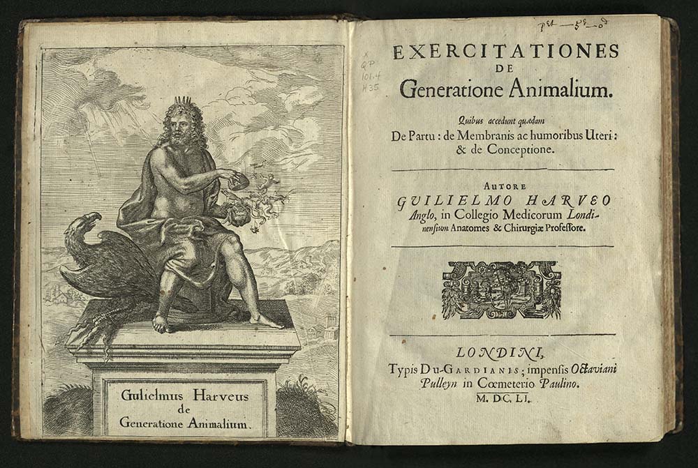
The English physician William Harvey discovered of the circulation of the blood. Harvey studied medicine at Cambridge and at Padua University (where Andreas Vesalius had taught a half century previously). Harvey was ahead of his time in the study of obstetrics. This work, founded upon empirical experimentation, contains the first original work on obstetrics published by an English author. Harvey included an anatomical description of the ovary of the hen, described the new-laid egg, then gave an account of the appearance seen on the successive days of incubation, and lastly described the process of hatching. He described the uterus of the doe, the process of impregnation, and the subsequent development of the fetus. Harvey insisted from these and other studies that all animals arise from ova. The theories proposed in this book were not to be proved until 1827. The engraved title-page depicts Jove holding a broken egg, out of which bursts all manner of animal and plant life, including a man, a reindeer, a grasshopper, and a dolphin.

QP29 D48 1662
Dutch physician and botanist Florentino Schuyl, the son of a pastor, lost his mother and two brothers to the plague in 1636. He began formal studies at the University of Utrecht, from which he received a degree in philosophy in 1639. He received his doctorate in medicine from the University of Leiden. His dissertation was on the spleen. He is best known for his translation into Latin of Rene Descartes’ work on physiology, Treatise of Man. Unfortunately, the copy he translated was not particularly accurate or complete. Schuyl, however, wrote an excellent introduction to Descartes’ ideas. Schuyl died in 1669 from plague. De Homine is the first in a series of works by French mathematician, philosopher, and physiologist Rene Descartes that set the agenda for later students of the distinction between mind and body. Descartes held back on the publication of this work after hearing of the condemnation of Galileo by the Inquisition. As a result, this work was published well after the author’s death.
QM21 H6 1669
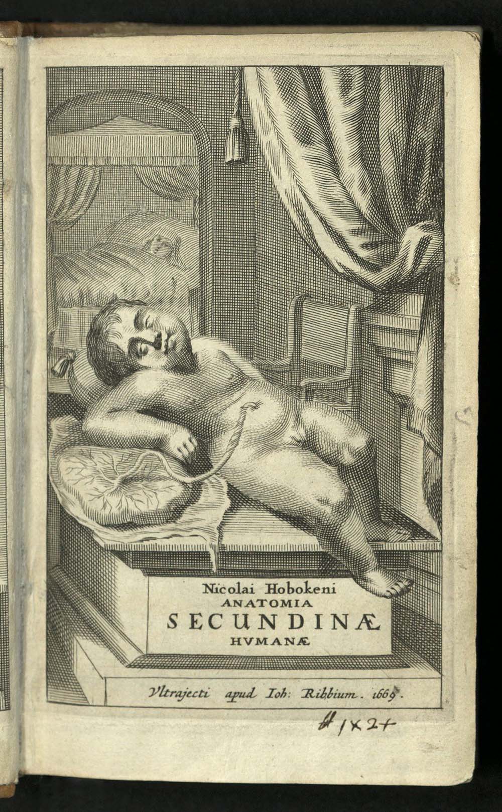
At the end of the seventeenth and into the first half of the eighteenth centuries, there was a renewed interest in the study comparative anatomy and physiology. Nicolaas Hoboken’s Anatomia was in the vanguard of these studies. Hoboken recieved a Doctorate of Medicine from Utrecht in 1662. The following year he was appointed professor of medicine at Steinfurt. From there he moved to Harderwijk where he became professor of medicine and mathematics. A second edition of Anatomia was printed in 1672 and a third in 1675. The book contains many woodcut illustrations within the text and seven large folding engraved plates.

QP301 B65 1680 v. 1, 2
Giovanni Borelli was trained in mathematics, but he also studied medicine, astronomy and geology. Among other things he investigated blood, Jupiter’s moons, and the movement of food through plants. At a time when Galileo was under attack, Borelli was sheltered from the Inquisition by Queen Christina of Sweden. Borelli corresponded with Galileo beginning in the 1640s, after Galileo’s house arrest. Borelli taught Marcelo Malpighi, who became a well-known anatomist. The two remained friends for life. In 1657, they founded a short-lived Italian scientific academy. Malpighi’s work inspired Borelli to study animal movement, a pursuit that would continue through the rest of his life. He would become known as the Father of Biomechanics, but Borelli died in poverty. His masterwork, De Motv animalivm (On the Movement of Animals), was published posthumously, one year after his death. Borelli followed Galileo’s model of geometrical analysis in mechanics as a way to explain biological phenomenon. He was the first to suggest that muscles contract with movement and that that contraction was enabled by chemical reaction in the muscle, and the first to recognize that forward motion entailed movement of a body’s center of gravity, and was then completed by the swinging of limbs to maintain balance. He compared the working of the heart to a piston.
QL804 B53 1681
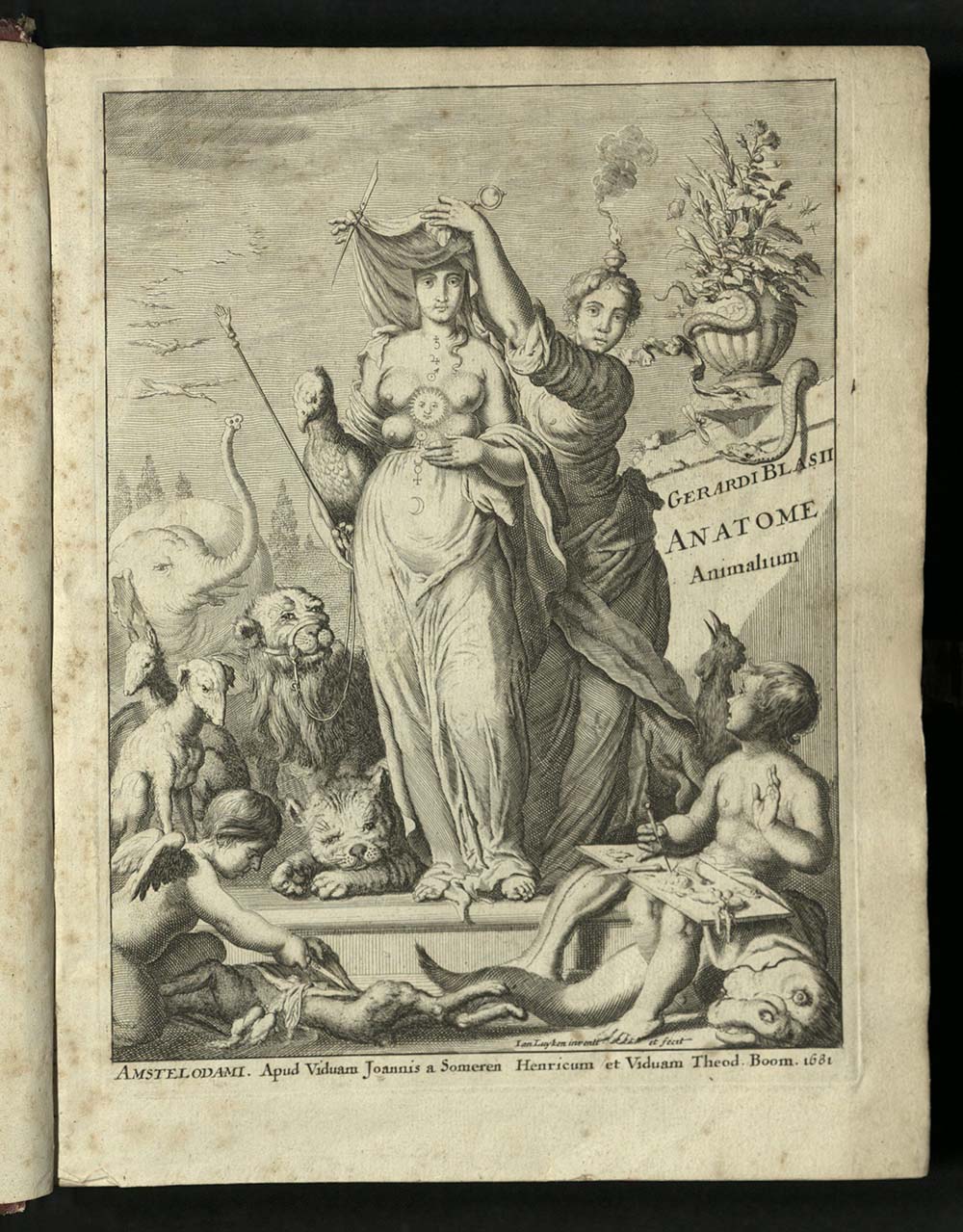
Gerard Blasius, a Dutch physician and anatomist combined some of his earlier writings with those by other scientists to put together this systematic treatise on comparative anatomy. Blasius’ father was an architect in Copenhagen, where Blasius began his studies. He practiced medicine in Amsterdam, where he developed his interest in anatomy, and in particular, comparative anatomy. He worked to widen the availability of both animal and human remains for study. He conducted dissections in hospital and in his home. Engraved plates depict animals and parts of animals accompanied by descriptions. The artists and engravers for this work are unknown, with the exception of Dutch illustrator and engraver Jan Luyken (1649-1712), who created the frontispiece.
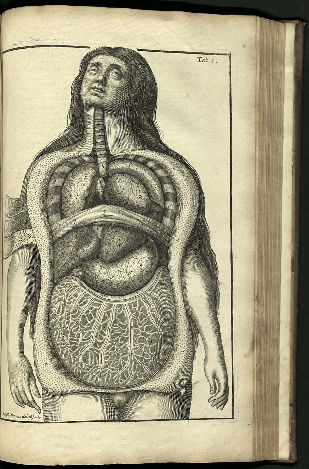
QL805 C65
The concept of comparing the brains of various animals and of individual human brains took shape early in the seventeenth century in England, influenced by the formation of the European scientific societies and their attempts to guide naturalist observations into a new systematics. This trend is typified in this lavishly illustrated work by Samuel Collins, an anatomist and president of the Royal College of Physicians. Collins wrote this in the vernacular, hoping for broad readership. The book had one of the largest collections of illustrations of the brain to date. The seventy-five copperplate engravings exemplified the direction of an “enlightened” scientific revolution. The illustrations, mostly signed by William Fairthorne, include observations on one hundred and fifteen species. There are early representations of the anatomy of the crab, the viper, and a silkworm. As a whole, the work represents the first attempt to produce a comprehensive treatise on comparative anatomy based on newly observed material. Collins’ focus, however, was to discover and explain the intricacies of man. Collins gratefully acknowledges the work of those who came before, including Marcello Malpighi. Title in red and black.
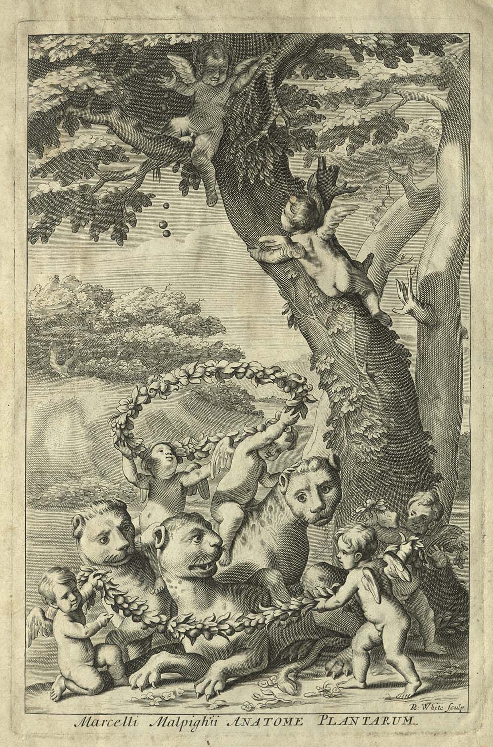
QH9 M2 1686
Marcello Malpighi, the founder of modern microscopic anatomy, made enduring contributions to the analysis and description of minute physical structures of living creatures from plants and insects to birds, frogs and humans. This collection of includes his work on the anatomy of plants; the embryonic development of the chick, the anatomy of the silkworm; and, his greatest contribution to science, the discovery of the existence of capillaries, which completed the chain of the circulation of blood postulated by William Harvey. Illustrated with engraved frontispiece and one hundred and sixteen full-page engraved plates, including seven small supplementary plates.
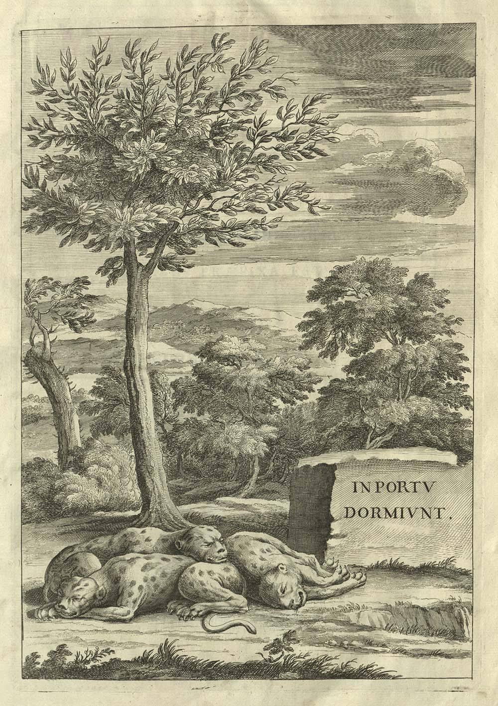
QH9 M3 1697
Marcello Malpighi was an Italian physician and anatomist. He taught at Pisa, Bologna, and Messina and was physician to Pope Innocent XII. While at Pisa, he discovered that the heart is composed of spiral fibers. He was admitted to the Royal Society as a fellow in 1661. Opera posthuma was put together by Malpighi specifically to be published after his death. It contains a series of manuscripts which he had not had time to complete and publish before his death. Malpighi worked to finish many of these manuscripts as he was dying, so that they could be published by the Royal Society, making his intention clear in a letter to the secretary of the Royal Society. The style in which he completed these pieces indicate that he felt he could be more forthright about his findings. His tone was noticiably more polemical than it had been in his other works. He defended all his previous work, addressing many of his critics, including Giovanni Borelli, who had criticized Malpighi’s work in his posthumously published De motu animalium (1680). He wrote that before using an instrument with greater magnification, one must first carry out observations under a simple microscope of lesser magnification. University of Utah copy bound with Malpighi’s D structura glandularum conglobatarum consimiliumque partium, epistola.
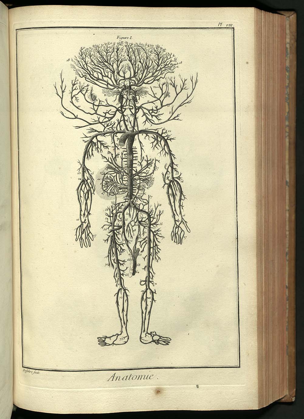
AE25 E53 plates vol. 1
Diderot said that the purpose of his Encyclopedie was “to change the common way of thinking.” Most of the finest thinkers of eighteenth-century France contributed to this monumental project, including such greats as Voltaire, Montesquieu, Rousseau, and the eminent mathematician Jean d’Alembert. What made the Encylopedie important was that it embodied a new approach to knowledge, based upon the new methods and theories of Francis Bacon and Isaac Newton. Diderot, and the other Encyclopedists who contributed to the project, refused to accept the authority of political power or religion in questions of the intellect or of art. Far from being a neutral compendium of useful knowledge, Diderot’s Encyclopedie, tantamount to a political manifesto,was an attempt on the part of radical French intellectuals to establish their own authority by virtue of their superior reason. They allowed nothing in the work that was not known through empirical study. Each volume became an immediate success. The French court, judiciary, and church, however, were all outraged by the project and what it stood for. In 1759, the seven volumes that had appeared to date were banned by the government and placed on the index by the Pope. For the next twenty years, Diderot carried on, writing countless small articles and many major ones, while dealing with the continuous attacks of critics. It is hard to imagine such a substantial work as an “underground” publication, but that, indeed, was what it was.
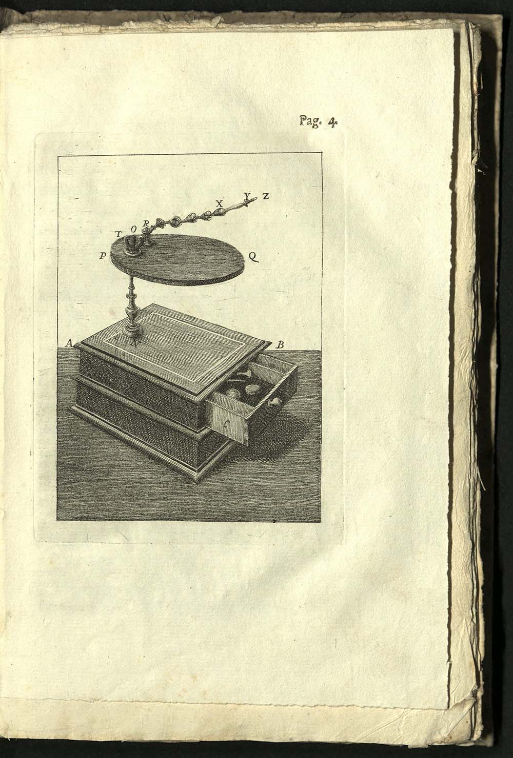
R128.7 B8
Although little is known of his life, from his own writings we know that Richard Brookes once practiced medicine in rural Surrey (see his Dedication in Art of Angling) and that sometime before 1762, he travelled to America and Africa (see his Preface in Natural History). Brookes also made some translations, most notably The Natural History of Chocolate (1724) from the French, Histoire Naturelle du Cacao et du Sucre (1719). His work went through several reprints. As late as 1834, Brookes’ books were listed in James Atkinson’s Medical Bibliography. Atkinson wrote that Brookes had “been a most industrious and intelligent practical author.” His were “books of considerable research and labour.”
QM21 H35
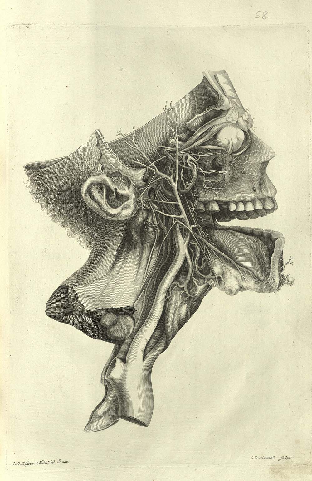
First issued as fascicles, Albrecht von Haller began this work in 1743, adding seven more pieces in 1845, 1847, 1849, 1852, 1853, 1854, and 1856. In 1756 the set was put together under the title, Icones anatomicae. The last four plates of the set, representing the arterial system of the whole body and consisting of two finished and two outline plates are twice as large as the others. The engravings in this work are exceptional. C.J. Rollinus, M.D., drew the plates in the first fascicle. Joel Paul Kaltehofer (d. 1777) engraved most of the plates. Other engravers included Daniel Heumann (1691-1759); Jacob van der Spyk of Leyden; and Carl Sedd of Amsterdam. Several of the plates represent, on a large scale, almost all of the arteries of the organs, with surrounding parts. Other representations include the diaphragm, the spinal cord, the uterus and the heart.
RB24 M67
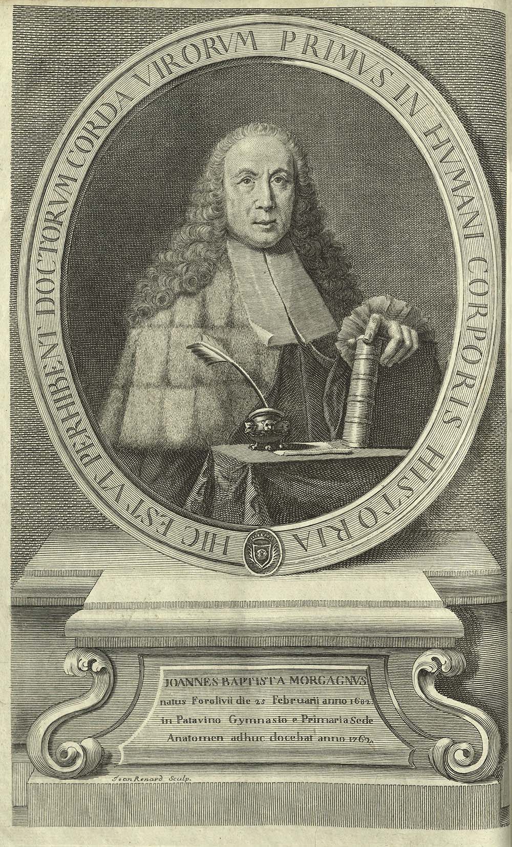
Giovanni Battista Morgagni was an Italian physician and anatomist, today recognized as the founder of anatomo-pathology. His first job was as an assistant to Antonio Maria Valsalva, who had been a pupil of Marcello Malphighi. De sedibus was published when Morgagni was seventy-nine years old. He wrote it over a twenty year period. In the form of a series of seventy letters, he gave detailed clinical descriptions of autopsy findings. The system he used to organize these findings provided a correlation between clinical symptoms with pathological findings, proving that postmortem examination was necessary for the study of a disease. For instance, Morgagni described ossificiation (calcification) of cardiac valves as an explanation for fatal heart conditions.
QP6 H32
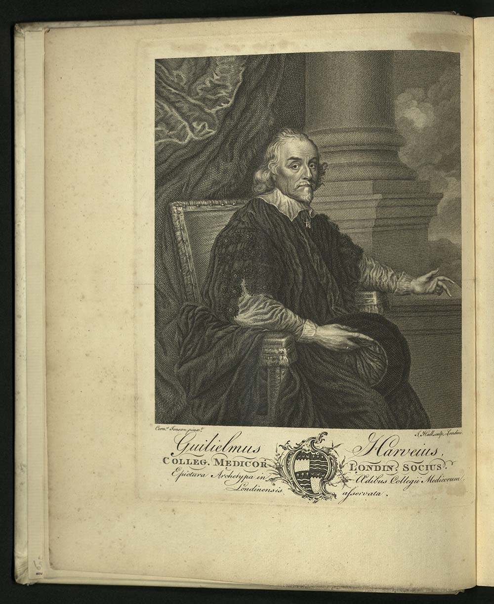
This collection of the works of the English anatomist and physician William Harvey, edited by Marke Akenside, was compiled under the direction of the Royal College of Physicians of London. The first edition of the collection was printed in 1737. The second edition is preferred by scholars and book collectors: it is far more accurate and complete. Included is the first printing of a biography of Harvey by Lawrence Harvey, an autopsy report by Thomas Parr, a number of unpublished letters, and the text of Harvey’s diploma from Padua. This edition contains nearly two hundred and fifty emendations to the 1628 Frankfurt edition of Harvey’s De Motu Cordis, and more than one hundred and fifty emendations to the 1649 Cambridge edition of his De sanguinis via the circulation. Edition of five hundred copies, of which one hundred and two were delivered to the crowned heads of Europe and one hundred and thirty-two copies to the College of Physicians. Records from the Bowyer print shop reveal that the production of this edition required “6 quires of writing royal for the plate, 19 reams 5 quires of writing royal and 745 reams of common royal.” University of Utah copy is a presentation copy from the London’s Royal College of Physicians to “Doctor Dickenson,” who delivered the Harveian Oration of 1891.

QP101 S67
Lazzaro Spallanzani was a Jesuit priest, biologist and physiologist. He was one of the first to disprove the theory of spontaneous generation, yet also investigated the ability of various animals to regenerate missing body parts. He proved that microorganisms could be killed with intense heat. In Dell’azione del cuore ne’ vasi sanguingi Spallanzani outlined his findings on the action of the heart upon the blood vessels. He demonstrated the rhythmic inequality of blood flow in the aorta and large vessels disappeared in medium and small arteries, becoming regular and uniform. The blood velocity in the venous system increased as the caliber of the vessels enlarged. Spallanzani dedicated this treatise to Albrecht von Haller.
QP101 S75
Lazzaro Spallanzani was the first to observe and demonstrate the passage of blood from the arteries into the veins through the capillaries in warm-blooded animals, using chicken embryos for his experiment.
QM21 C97 1790
William Cruikshank was a British chemist and anatomist. In 1771, he became assistant to William Hunter. He then became partner in the Great Windmill Street School, later named the Hunterian Medical School. In 1797, the same year he was elected a Fellow of the Royal Society, he first demonstrated that a particular crystallizable substance exists in urine and is precipitated from it by nitric acid. In 1800 he identified carbon monoxide as compound containing carbon and oxygen. That same year, he used chlorine to purify water. In his Anatomy of the Absorbing Vessels, first published in 1786, he traced the lymphatic vessels through the human body, as well as in numerous animals. This work was later translated into French, German and Italian. Cruikshank was the attending physician at the deathbed of Dr. Samuel Johnson (1709-1784), who once called him a “sweet-blooded man,” referring to his complacent disposition. Cruikshank, however, died at an early age of apoplexy brought on by alcoholism. The second edition of Anatomy is illustrated with five plates, three of which are printed in red, two of which are folding.

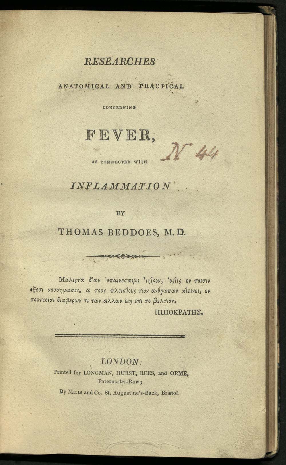
RB129 B43 1807
While at Pembroke College, Oxford, studying medicine, Thomas Beddoes became interested in chemistry and also studied modern languages. He translated or edited translations of the works of Lazzaro Spallanzani (1729-1799), among others. Between 1784 and 1788, Beddoes visited Edinburgh and France where he met Antoine Lavoisier (1743-1794). In 1788, Beddoes was appointed reader in chemistry at Oxford, attracting the largest audiences since the 13th century. Beddoes was particularly concerned with tuberculosis. He treated patients by having cows breathe on them, based on his observance that butchers appeared to avoid the disease. In spite of this, he was skeptical when Edward Jenner (1749-1823) began using a cow-derived vaccination for smallpox a few years later. In Researches Anatomical, Beddoes contended that fever and inflammation of organs and tissues are related by one causing the other. He extensively treated the subject of morbid anatomy and described the conditions of various organs in their post-mortem state. The appendix contains accounts of various ailments and their cures, including a successful treatment of rabies, “…by copious bleeding and mercury.” The announcement came in the form of two letters from Dr. Robert Burton of Bent, Virginia to Dr. Benjamin Rush of Philadelphia. University of Utah copy has the inscription on the verso of the title-page: “To the Library of the Royal West London Infirmary presented by B. Golding M.D. first director of the Charity at its establishment Anno 1818.”
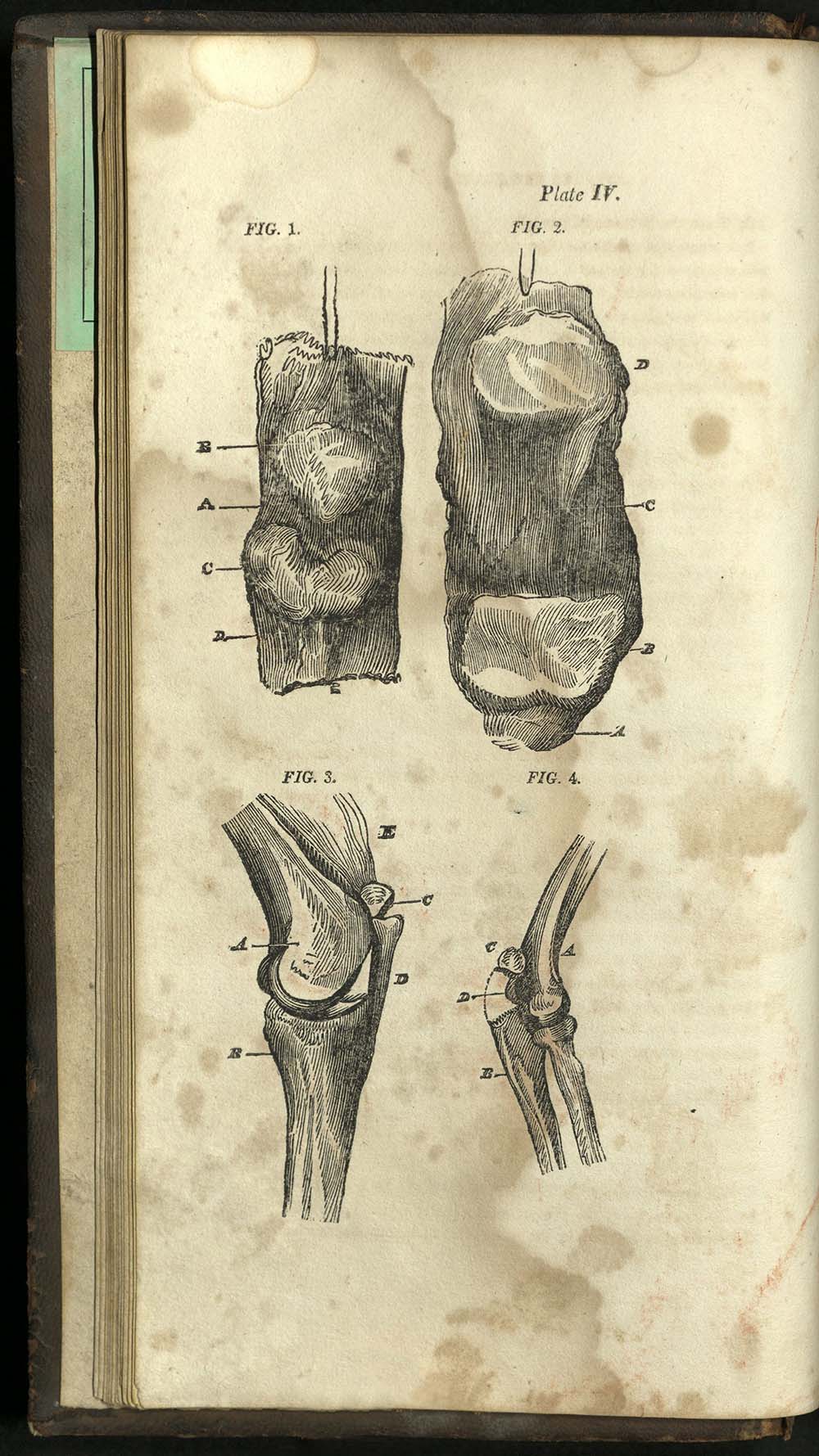
RD32 B4
Charles Bell, born in Edinburgh, was a surgeon who served with the British Army at the Battle of Waterloo. In 1767, Bell took over the school of anatomy in Great Windmill Street, founded by William Hunter that same year. A professor of surgery at the University of Edinburgh, he is best known for his medical illustration and for his pioneering study of the human nervous system. He accurately described the thoracic nerve, or, “Bell’s nerve;” he discovered that a lesion of the seventh facial nerve causes paralysis, “Bell’s palsy;” and he demonstrated the function of spinal nerves, i.e. that motor function relates to anterior roots and sensory function relates to dorsal roots, the “Bell-Magendie law.” In 1807, Bell published his System of Operative Surgery, where he described surgical operations based on personal experience. As an appendix to this work, Bell added an essay on gun-shot wounds, based on knowledge he acquired as a surgeon for the wounded of Waterloo.

QM11 B87 1840
Surgeon and lecturer of human anatomy at the University of Ghent, where he received his degree in medicine, Adolphe Burggraeve also designed a clean, affordable housing for workers in the city. He was active in civil and political affairs, particularly concerned with the poor of Ghent. He was the author of many works, including two books on Andreas Vesalius – his life, and his impact on medicine. University of Utah copy gift of Mrs. Thomas Kearns.
RB24 L42
In the early nineteenth century, Paris was the center of Europe’s medical world. A transition from speculative medicine to medicine based on rational observation, begun more than a century earlier during the Enlightenment, was taking place. Hermann Lebert studied medicine in Berlin, Zürich, and Paris, where he also practiced medicine. He moved to Paris in 1846, where his work fighting a cholera epidemic won him the cross of the Legion d’honneur. In 1852, he went to Zürich and in 1859, he returned to his hometown of Breslau. He was a prolific writer in both German and French. He was one of the first anatomists of the nineteenth century to use the microscope for pathological diagnostic studies. In his research, he distinguished between tuberculosis and cancer. In Traité D'Anatomie Pathologique, Générale et Spéciale, Lebert was one of the first to describe premalignant polyps of the colon, rectum, and stomach. Traité D'Anatomie Pathologique was the most comprehensive illustrated medical dictionary to date, containing clinical material from the best-regarded physicians of France. The illustrations are steel-face copper engravings printed in color using multiple runs by the most renowned publishing house of the time.
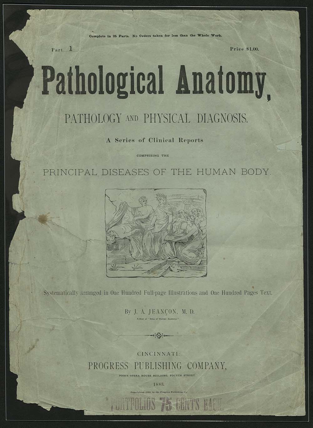
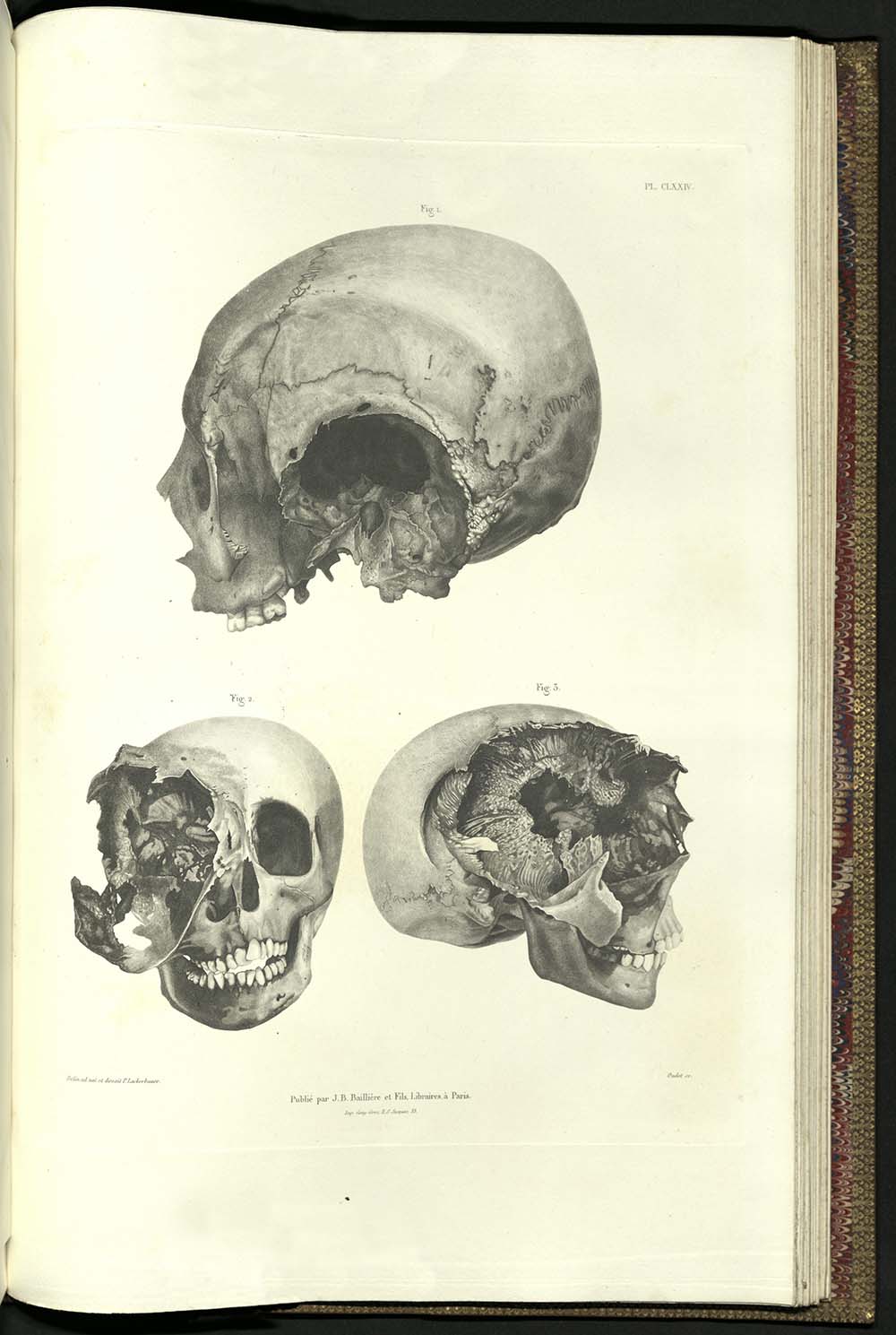
RB33 J4
Born in Cabrai, France, John Allard Jeancon was educated in Paris, Turin, and Berlin before being graduated with a medical degree from the Royal College of Surgeons at Edinburgh in 1854. He immigrated to the United States, where, in 1854, he began teaching at the Eclectic Medical Institute (EMI) in Cincinnati, Ohio. He served as a surgeon in the 32nd Regiment of Indiana Volunteers during the Civil War. While serving, he, along with other surgeons and the United States Surgeon-General, protested the indiscriminate use of mercury as a drug by the army, eventually leading to its ban. After the war, he returned to EMI until he retired in 1891. Pathological Anatomy contains case studies of patients, with full-color plates of pathological ailments. Issued in twenty-five parts, sold as a whole.
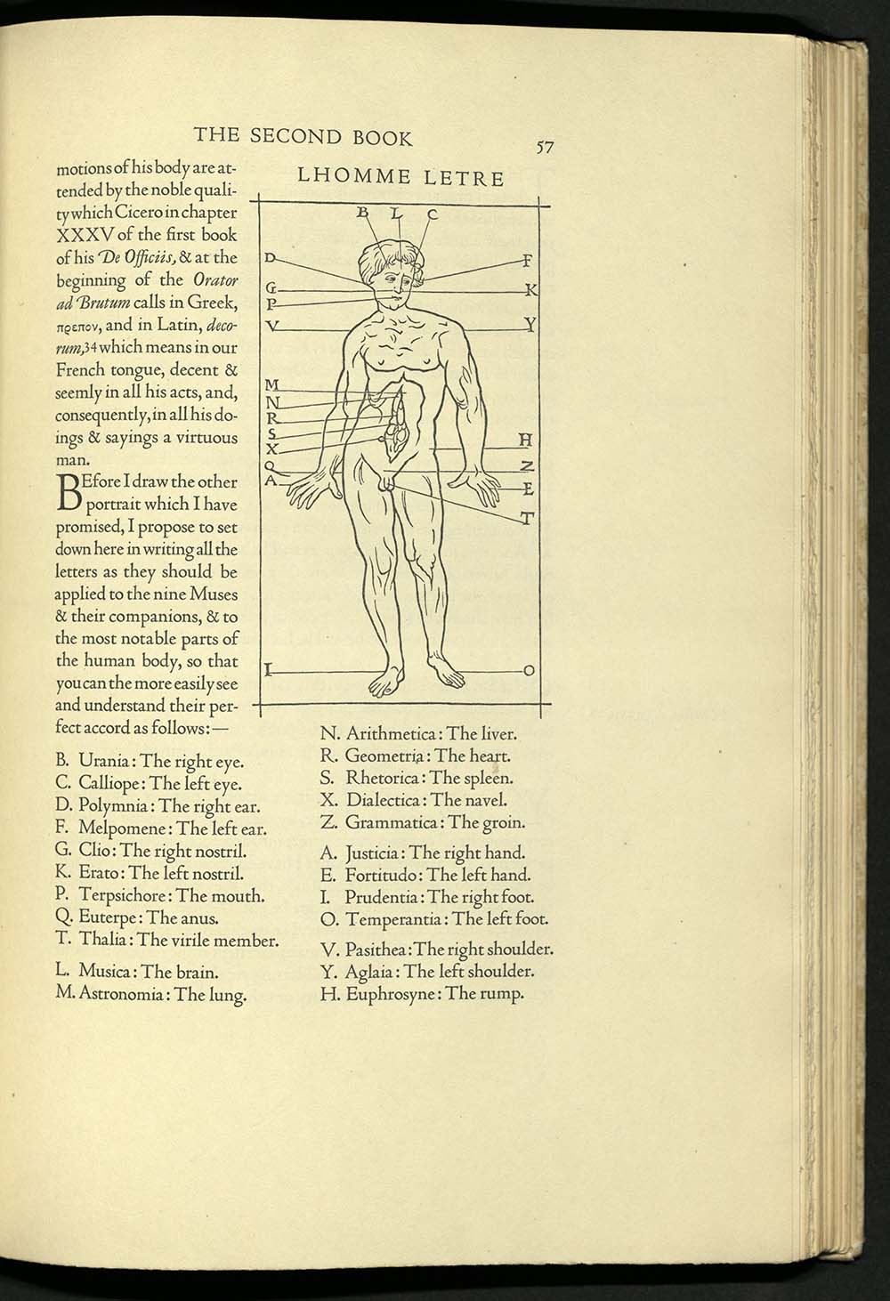
NK3615 T62 1927
This is the first edition in English of Geofroy Tory’s famous work on Roman letterforms, first published in Paris in 1529. Tory, one of the major printers in Paris during the first third of the sixteenth century, wrote and printed this theoretical treatise on the design of Roman capital letters in 1529. In 1531 he was named Imprimeur du roi. Tory developed a method of drawing letters with geometrical aids and then related their proportions to the human body. The English edition was printed for the Grolier Club at The Printing House of William Edwin Rudge and designed by Bruce Rogers, one of the premier American typographers of the twentieth century. The typeface is Centaur, designed by Rogers and based on traditional Venetian typefaces first developed around 1465 by master printer Nicholas Jensen. Rogers redrew all the original specimens and illustrations, thereby omitting the imperfections resulting from the over-inking and poor printing of the Paris original. This book was one of Rogers’ own favorites. Printed on American woven rag paper, it was bound in half vellum, lettered in gold, and covered with a paper in a pattern of alternating fleurs-de-lys and thistles, also designed by Rogers. The University of Utah’s copy is signed by Bruce Rogers.
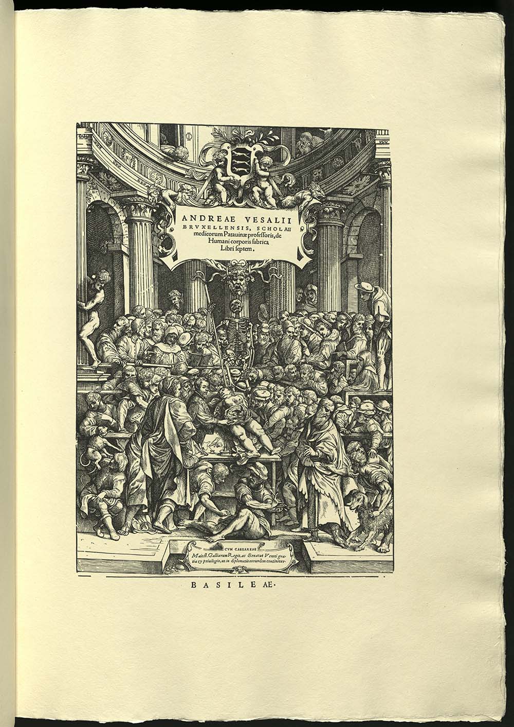
QM25 V4 1934 oversize
The woodblocks for Vesalius’ anatomical work were one of the great discoveries – and losses – of the book world. Upon the death of the printer of the first and second editions of Vesalius’ “Anatomy” the woodblocks were given to Jerome Froben’s son, grandson of Johann Froben, under whom printer Johannes Oporinus apprenticed. They remained with the Froben family printing business until it closed shop with the third generation in 1603. The blocks were transferred from Basel to Augsburg, where a few of them were published in the early eighteenth century. Later that century many of the blocks appeared in work published in Ingolstadt. In 1800, Ingolstadt was captured by the French, and the blocks were evacuated to Landshut in Bavaria. In 1826 they were transferred to the University of Munich. There, they went unrecognized until they were discovered in an inventory taken in 1893. In 1932 the two hundred and twenty-seven surviving blocks were rediscovered. Willy Weigand, who worked on this project, wrote, "the woodblocks were as though they had been cut today.” For this edition the woodblocks preserved in Munich were joined by the original woodblock for the title-page of the 1555 Fabrica from the Library of the University of Louvain. The publication of this edition was a formidable task, designed to demonstrate to a new audience the beauty of the original edition. The publication so soon after the rediscovery was particularly fortunate. The blocks were destroyed during WWII. Edition of six hundred and fifteen copies.
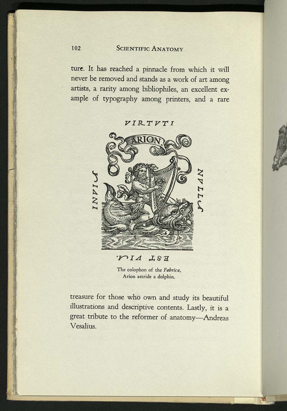
PS3535 U447 Q8 1944
Nolie Mumey gives tribute to Andreas Vesalius, celebrating the four hundredth year of the publication of “The Fabrica” with a sketch of its content. Surgeon Nolie Mumey was graduated from the University of Arkansas Medical School in 1916. He went on to earn a Master of Science degree from the University of Pennsylvania and a BA and MA from the University of Denver. A twentieth-century Renaissance man, he was a poet, a silversmith, aviator, carpenter, woodcarver, artists, and inventor. He had an extensive collection of books and artifacts of the American West and its history. He wrote numerous books on Western history and medical history. Illustrated with line art reproductions and one folding plate. Edition of one hundred and fifty copies. University of Utah copy is no. 22, signed by the author. Gift of S. Lyman Tyler.

N7433.4 A64 A63 1990z
Two-sided pamphlet stitched into a dos-a-dos structure with map sheets that unfold perpendicular to each other. Printed by Lori Spencer on a Heidelberg Offset Press. The font is Olaus Magnus. The paper is white Mohawk Recycles 80LB text. Edition of two hundred and nine copies. University of Utah copy is no. 16.
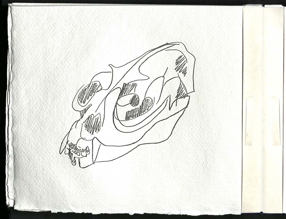
N7433.4 B884 B66 1992
A group of twenty poems inspired by a series of bone and skull drawings by Ruth Fine, eighteen of which are included in this edition. The typeface on forty french-folded pages is 10 point Gill Sans Light printed on Barcham Green Royal Watercolour society paper. Bound in a woven non-adhesive structure on MacGregor-Vinzani calendered ivory abaca paper. Housed in two slipcases; one of Barcham Green’s Renaissance IV made from old British Mailbags; the other is drum vellum. Both slipcases are constructed without glue. The entire structure designed and executed by Claire Van Vliet. Edition of one hundred and fifty copies, numbered and signed by the author, illustrator, and printer. University of Utah copy is no. 24.
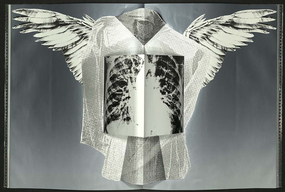
N7433.4 D675 M4 1993
Texts by Freud and others. First mounted as a computerized slide and sound installation in the Brooklyn Bridge Anchorage in 1990. Presented as a radio piece in the 1991 New American radio series, then as an essay in the summer of 1992 edition of the N.Y.U. drama journal. Using transparent and opaque metallic papers (including a three-dimensional centerfold pop-up), this book’s many layers create a rich and densely visual reading experience. Printed offset in several shades of metallic ink by Lori Spencer at the Borowsky Center for Publication Arts. Bound in perforated metal boards with screen mesh and iridescent plastic fly leaves by Daniel Kelm and staff at the Wide Awake Garage. Issued in slipcase by Jill Jevne covered with silver leaf. Edition of sixty copies, ten hors de commerce. University of Utah copy is no. 24.

PS3560 O4557 A62 1997
Twenty detailed and technically accurate anatomical poems. Alice Jones practiced medicine before she became a psychoanalyst. The poems were originally published in various journals and reviews including the JAMA, the New England Journal of Medicine, the UCSF Poetry Anthology and the Lancet. Illustrated with eight multi-colored reduction linoleum block prints representing microscopic views of the body. Titles printed in scarlet. Designed and printed by Asa Peavy. The type is Gill Sans Medum, cast by Michael & Winifred Bixler, printed on Rives Lightweight paper using a Vandercook SP-15 press. Edition of one hundred copies, eighty-five of which were bound by Coriander Reisbord in blood red pastepaper boards with a transparent vellum spine. The remainder are offered in full vellum or in sheets.
N7433.4 C865 A53 2004 Oversize
Fasciculus medicinae (1495) was the first printed book with anatomical illustrations – some of which are reproduced here. A hologram on the cover of this book is of an early-eighteenth-century brass lancet, or scalpel. Because of the book’s massive size the text was digitally composed using Adobe Poetica, a typeface designed to reflect the humanist handwriting style of the Renaissance. The companion typeface is Minion, designed by Robert Slimbach at Adobe, a clean, streamlined, highly functional roman. Printed from eighty-five photopolymer plates made by Boxcar Press. Cotton paper handmade by John and Kathy Koller. Edited and letterpress printed by Robin Price with assistance from Anne Thompson. The concertina binding co-designed by Daniel E. Kelm, Robin Price and Joyce Cutler-Shaw. Issued in custom-made, burnished stainless steel boxes fabricated and engraved by Pina Zangaro. Edition of fifty copies. University of Utah copy is no. 23.

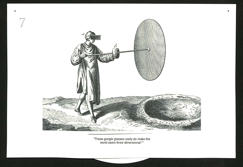
N7433.4 H62 D53 2014
Ten volvelles contained in a box covered with the image of the spines of the Diderot Encyclopédie, which described with articles and depicted with three thousand engravings many of the mechanical processes that would help transform the world. Text on volvelles includes original captions in French, with alternative captions in English. From the artist: “I made copies of several of the engraved images…and circulated them to friends soliciting their suggestions for contemporary captions. I am indebted to these collaborators: Vince Ricci, Alice Shaw, Michael O’Shea, Judy O’Shea, Alisa Golden, Bob Breihan, Nance O’Banion, and Sandra Hobson.” Designed and assembled by Charles Hobson with the assistance of Alice Shaw. Edition of twelve copies.
QM21 V413 2014
From the publisher: “This title recreates the masterpiece of science and art for the first time in a way that is understandable to 21st century readers who do not have any knowledge of Latin. The texts of both the 1543 and the 1555 editions have been translated with the utmost care by Northwestern University Professors Emeritus Daniel H. Garrison and Malcolm H. Hast, a task they completed in over twenty years of painstaking and dedicated work. Annotations give the reader keen insight into just how innovative ‘De humani corporis fabrica’ was, and high-resolution digital scans of the almost three hundred woodcuts provide the images with a sharpness they never had before.”